
Case 19 – Surf boarded
October 17, 2018
Case 21 – Checkhamate
October 17, 2018Case 20 – Tib of the iceburg

Case 20
29 year old presents with right ankle pain — especially with running and jumping, cycling is fine. 3 years ago the patient inverted his ankle while playing cricket and the pain has been experienced ever since. Describes pain as across anterior and posterior ankle joint. Has previously had an MRI showing osteoarthritic findings in the ankle but no fracture. No previous surgeries.
On exam, slight swelling, no deformity or ecchymosis. Loss of range of motion and ankle effusion. No signs of ligamentous instability — anterior draw test and talar tilt test negative.
Assessment: Given age and no prior injuries to 2015 — recommended a repeat MRI.
Imaging:
Figure 1: Coronal view MRI of the right ankle showing an osteochondral defect in the posterior medial tibia.
Figure 2: T1 weighted sagittal MRI of the right ankle. An osteochondral defect can be seen in the posterior medial tibia.
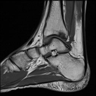
Figure 3: T2 weighted sagittal MRI of the right ankle showing an osteochondral defect in the
posterior medial tibia along with an area of bony edema posterior to the tibia.

Discussion:
Osteochondral defects usually involves the articular hyaline cartilage and the subchondral bone. The defect may be caused by cartilage damage due to shearing stresses while the subchondral bone is intact, or by a high-impact force causing bone contusion. Pathology can range from a simple contusion of the cartilage and subchondral bone to a fracture of cartilage alone or cartilage and underlying subchondral bone. Osteochondral defects of the ankle can occur from a lesion caused by single or multiple traumatic events, leading to partial (stable) to complete detachment of the fragment (unstable). Commonly, ankle sprains, particularly inversion injuries, can be the cause. These injuries usually cause deep ankle pain during weight bearing. Impaired function, limited range of motion, stiffness, swelling and joint effusion may also be present. If symptoms have not resolved within 4-6 weeks then an osteochondral defect should be suspected. The talar dome is the most common area where ankle osteochondral defects occur. The defect may heal, remain asymptotic or progress to an subchondral cyst. Plain radiographs are a common in the initial evaluation of osteochondral defects but the defect may not be visible. MRI and CT have been shown to be more accurate.
When assessing an ankle injury, inquire about the exact mechanism of injury, ability to walk after injury and previous injuries or surgeries. On exam, check for signs of swelling, tenderness and ecchymosis, which are indicative of acute ankle injury. Before extended physical exam, use the Ottawa ankle rules to evaluate the need for radiography. The rules state that ankle radiography is needed if there is bone tenderness over the posterior edge of the medial or lateral malleolus or an inability to walk four step immediately after injury and in the physician’s office. Foot radiography is required if there is bone tenderness at the base of the fifth metatarsal or at the navicular bone or if there is an inability to walk four steps immediately after the injury and in the physician’s office.
Diagnosis: Osteochondral defect in the posterior medial tibia.
Treatment:
Given the length of injury and multiple rounds of physiotherapy, refer for surgical consultation. Surgical treatment may include excision of the lesion, excision, excision combined with filling of the defect with autogenous cancellous bone graft (microfracturing), fixation techniques, or osteochondral transplantation (OATS – osteochondral autograft transfer system).
Author: Lucas Nguyen October 17, 2018 (PRND)
References:
- Van Dijk, C. N., Reilingh, M. L., Zengerink, M., & van Bergen, C. J. A. (2010). Osteochondral defects in the ankle: why painful? Knee Surgery, Sports Traumatology, Arthroscopy, 18(5), 570– http://doi.org/10.1007/s00167-010-1064-x
- Badekas, T., Takvorian, M., & Souras, N. (2013). Treatment principles for osteochondral lesions in foot and ankle. International Orthopaedics, 37(9), 1697– http://doi.org/10.1007/s00264-013-2076-1
- Van Bergen, C. J., Gerards, R. M., Opdam, K. T., Terra, M. P., & Kerkhoffs, G. M. (2015). Diagnosing, planning and evaluating osteochondral ankle defects with imaging modalities. World Journal of Orthopedics, 6(11), 944– http://doi.org/10.5312/wjo.v6.i11.944


